
Figure 2.
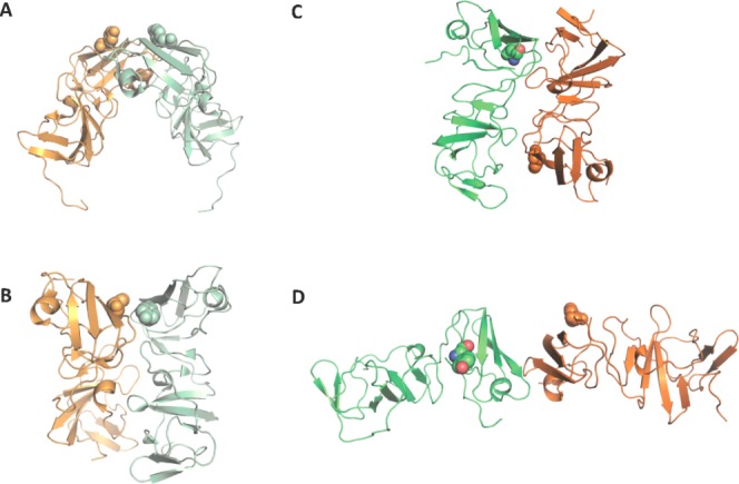
Genotype 1b and 1a NS5A domain 1 crystal structure dimers in ribbon representation. The side chain of tyrosine 93, a major resistant mutation, is displayed as spheres. Monomers are shown in orange and green, and coloured in pastel for genotype 1b dimers. (A) The first crystal structure dimer31 forms a potential RNA-binding pocket. (B) The second genotype 1b dimer32 are juxtaposed in parallel to form an extensive interface. (C) Monomers A and B from the genotype 1a NS5A domain 1 crystal structure share the same interface as the Love et al. dimer shown in B albeit with the monomers arranged in an antiparallel fashion. Use this link to access the interactive version of this figure. (D) Monomers C and D form a N-terminal, head to head dimer. Use this link to access the interactive version of this figure.