
Figure 6.
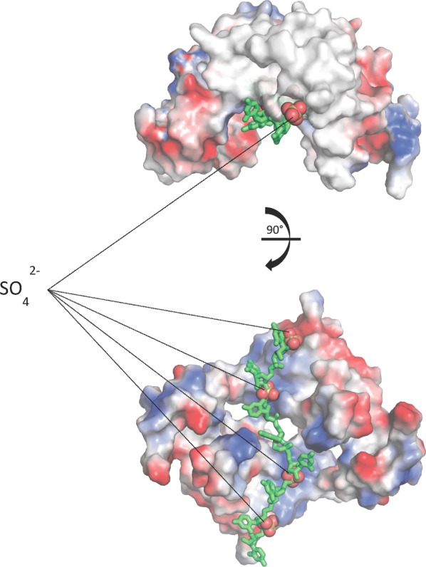
Viral RNA approximately modeled into NS5A domain 1 dimer. A structural alignment of monomer A and its associated sulfates from our genotype 1a crystal structure with each monomer in the Tellinghuisen et al. dimer places the sulfates in the dimer's RNA-binding groove. The sulfates may be mimicking an RNA phosphate backbone allowing the modeling of viral RNA into the dimer. Use this link to access the interactive version of this figure.