
Figure 2.
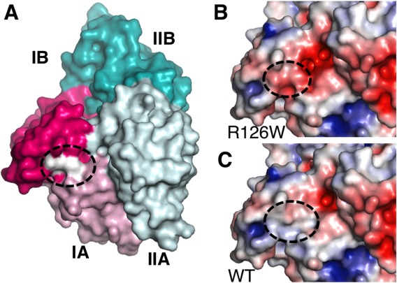
Location and effect of the Parkinson's disease mutation R126W. (A) Location of R126 mapped onto the Mortalin-NBD surface. NBD subdomains are colored and labeled as in Figure 1. Location of R126 and the nearby D130 and Q201 residues are shown as a white patch and marked by a dashed oval. (B) Surface electrostatic potential of the R126W mutant calculated using APBS,62 within a range of −5(red) to +5kT (blue). The dashed oval marks the location of W126, D130, and Q201. (C) Surface electrostatic potential of wild-type Mortalin-NBD, colored and marked as in panel (B). Use this link to access the interactive version of this figure.