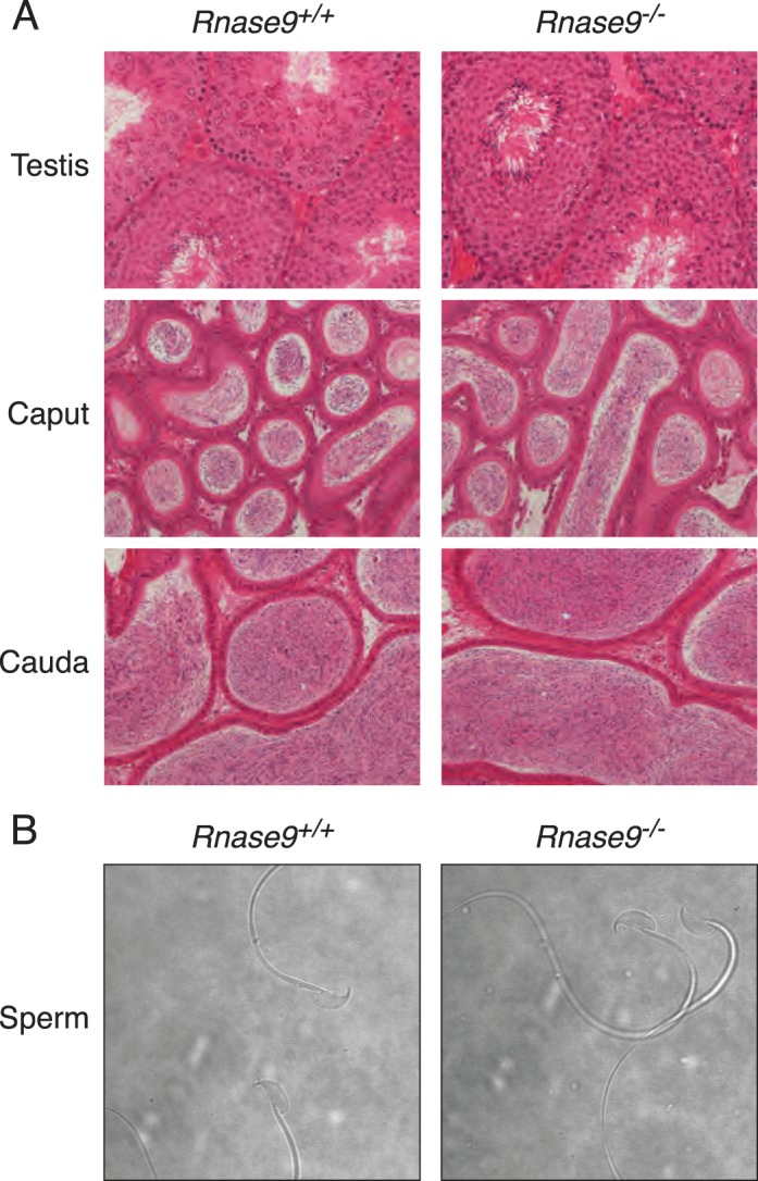
FIG. 2.

Tissue histology and sperm cytology. A) Hematoxylin-eosin stained sections of tissues. Testes (top panels), caput (middle panels), and cauda epididymides (bottom panels). Images are representative of three different sexually mature, age-matched Rnase9+/+ (left panels) and Rnase9−/− (right panels) mice. Original magnification ×20. B) Phase contrast images of caudal epididymal sperm. Images are representative of three different sexually mature, age-matched Rnase9+/+ (left panel) and Rnase9−/− (right panel) mice. Original magnification ×60.
