FIG. 4.
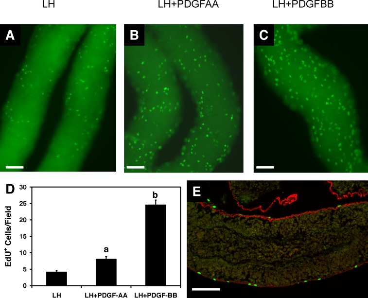
Cell proliferation in response to PDGF-AA and PDGF-BB during the first week of culture. Micrographs showing EdU-positive cells after the tubules were cultured for 5 days with LH (A), LH plus PDGF-AA (B), or LH plus PDGF-BB (C). D) The numbers of EdU-positive cells per tubule length for groups A through C were determined. Different letters indicate significant differences. Error bars are the mean (SEM). E) Click-iT EdU staining (green) on the surface of the tubules. The basement membrane is immunofluorescently labeled with anti-laminin antibody (red). Bar = 100 μm.

