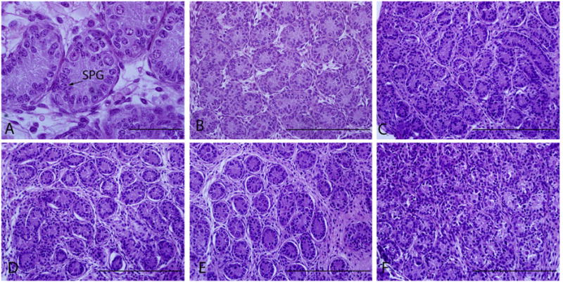
Fig. 2.

Morphology of fresh and frozen–thawed pre-grafted testicular tissue. Fresh control (A and B), cryopreserved neonatal testes with DMSO (C), PrOH (D), glycerol (E), and EG (F). Original magnification of A is ×100 and scale bar represents 50 μm. Original magnification of B–F are ×40 and scale bars represent 200 μm. Arrow points spermatogonia (SPG).
