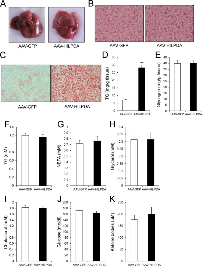
FIGURE 5.
AAV-mediated HILPDA overexpression induces hepatic steatosis. A, gross morphology of livers from AAV-GFP and AAV-HILPDA animals 4 weeks after injection of viruses. Animals were sacrificed under fed conditions. B, representative H&E staining of livers from AAV-GFP and AAV-HILPDA animals. C, Oil Red O staining of liver sections from AAV-GFP and AAV-HILPDA animals. D, hepatic TG content determined 4 weeks postinjection of AAVs (n = 8 animals/group). E, hepatic glycogen levels in AAV-GFP and AAV-HILPDA animals (n = 8 animals/group). F–K, plasma TG (F), NEFA (G), glycerol (H), cholesterol (I), glucose (J), and ketone body (K) levels in AAV-GFP and AAV-HILPDA animals 4 weeks postinjection of respective viruses (n = 7–8 animals/group). Data are mean ± S.E. (error bars). Asterisks, significant difference according to Student's t test (**, p < 0.01).

