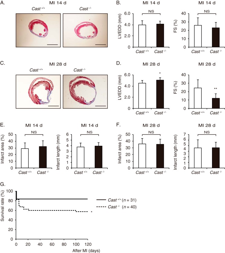
FIGURE 2.
LV remodeling after MI in Cast−/− and Cast+/+ mice. A, Masson's trichrome staining of Cast−/− and Cast+/+ hearts at 14 days after MI. Scale bars, 2 mm. B, echocardiographic parameters of Cast−/− and Cast+/+ mice at 14 days after MI. C, Masson's trichrome staining of Cast−/− and Cast+/+ hearts at 28 days after MI. Scale bars, 2 mm. D, echocardiographic parameters of Cast−/− and Cast+/+ mice at 28 days after MI. E and F, infarct area (light panels) and infarct length (right panels) of Cast−/− and Cast+/+ hearts at 14 days (E) and 28 days (F) after MI. G, Kaplan-Meier survival curves of Cast+/+ (n = 31) and Cast−/− mice (n = 40) after MI. LVEDD, LV end-diastolic dimension; FS, fractional shortening. Values represent the mean ± S.E. of data from 10 mice in each group. NS, not significant. *, p < 0.05; **, p < 0.01 versus Cast+/+ mice.

