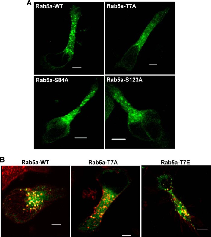
FIGURE 5.
Subcellular localization of Rab5a and its targeting to T-cell endosomal compartments. A, shown are representative confocal images of HuT-78 T-cells expressing GFP conjugated wild-type Rab5a (Rab5a-WT) or mutant Rab5a-T7A, Rab5a-S84A, or Rab5a-S123A after stimulation by incubating on an anti-LFA-1 coated surface for 1 h. B, HuT-78 cells were transfected with plasmid constructs expressing GFP conjugated wild-type Rab5a (Rab5a-WT), mutant Rab5a-T7A, or Rab5a-T7E and stimulated via LFA-1 as above. Cells were treated with AlexaFluor® 568-labeled transferrin (red) to allow internalization for 10 min and imaged. At least 20 microscopic fields per slide prepared from three independent experiments were visualized, and a representative data is shown. Scale bar, 5 μm.

