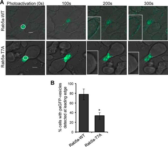
FIGURE 7.
Photoactivated Rab5a-paGFP- but not Rab5a-T7A-paGFP-associated vesicles redistribute from the centrosomal region to the leading edge of T-cells during migration. A, HuT-78 T-cells expressing wild-type Rab5a-paGFP (Rab5a-WT) or mutant Rab5a-T7A-paGFP (Rab5a-T7A) were stimulated to migrate on anti-LFA-1-coated coverslips and imaged by confocal microscopy. Photoactivation was performed within the indicated white circle around the centrosomal region of the cell. The leading edges of the migrating cell at 200 and 300 s post-photoactivation are shown enlarged in the insets. B, percentage of cells (±S.E.) with Rab5a-WT- or Rab5a-T7A-associated vesicles detected at the leading edge of migrating cells was plotted. n = 19 for Rab5a-WT and 25 for Rab5a-T7A. *, p < 0.01. Results are representative of three independent experiments.

