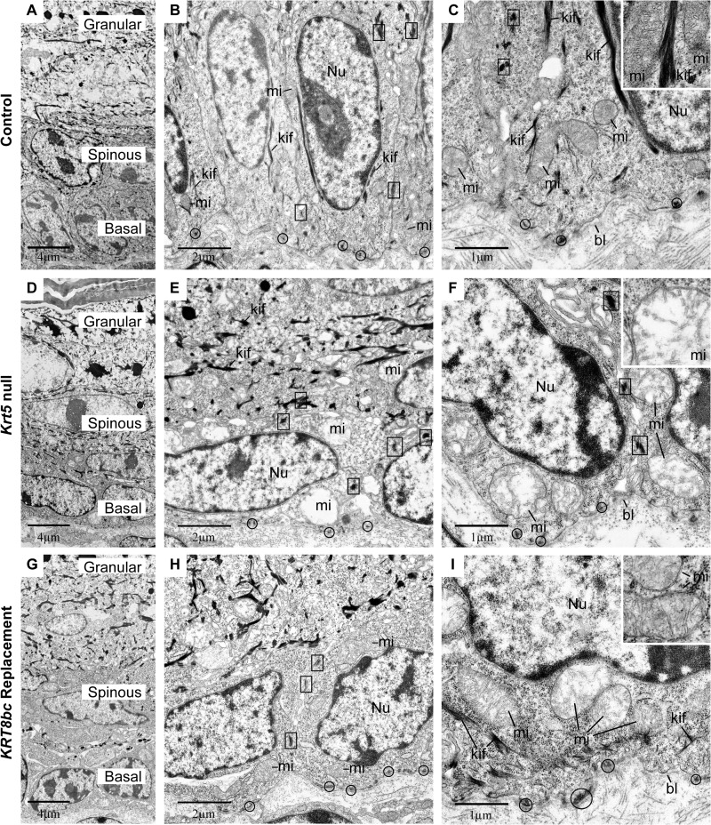
FIGURE 4.
Ultrastructural analysis of E18.5 epidermis in situ. Krt5+/+ (Control; A–C), Krt5−/− (Krt5 null; D–F), and KRT8bcTg/−Krt5−/− (KRT8bc Replacement; G–I) mouse epidermis at E18.5 was analyzed by routine transmission electron microscopy of thin sections. A, D, and G provide low magnification surveys of the living layers of epidermis (basal, spinous, and granular), whereas all other panels provide details of basal keratinocytes. bl, basal lamina; kif, keratin intermediate filament bundles; mi, mitochondria; Nu, nucleus. Examples of hemidesmosomes and desmosomes are circled and boxed, respectively, and representative mitochondria are detailed in the insets. Bars, 4 μm in A, D, and G; 2 μm in B, E, and H; and 1 μm in C, F, and I.

