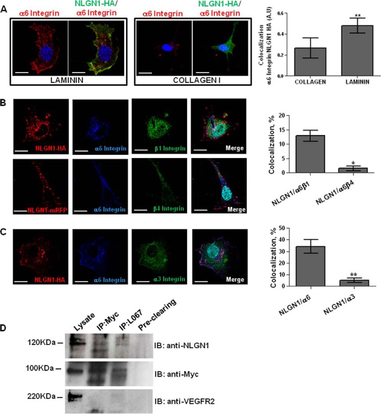
FIGURE 5.
NLGN1 and α6 integrin: colocalization and co-immunoprecipitation in ECs. A–C, confocal microscopy analysis performed on ECs transfected with mouse NLGN1-HA (mNLGN1-HA) or NLGN1-mRFP and plated 2 h on laminin (10 μg/ml) or collagen I (10 μg/ml). A, NLGN1 and α6 integrin colocalize on laminin. ECs were immunostained with anti-HA (green) and anti-α6 integrin (red) antibodies. The images are representative of 1 of 3 reproducible experiments. Scale bar: 10 μm. The graph shows the colocalization index ± S.E. (n = 3, total cells analyzed n = 10), unpaired Student's t test, two-tailed (**, p = 0.0011) for laminin versus collagen I. B, NLGN1 and α6β1 integrin co-localize in ECs plated on laminin. ECs were immunostained with anti-HA (red), anti-α6 integrin (blue) in combination with anti-β1 integrin (green, upper panel) or anti-β4 integrin (green, lower panel) antibodies. The images are representative of 1 of 5 reproducible experiments. Scale bar: 10 μm. The graph shows the percentage of colocalization between NLGN1/α6/β1 integrin and NLGN1/α6/β4 integrin ± S.E. (n = 3, total cells analyzed n = 10), unpaired Student's t test, two-tailed (*, p = 0.023). C, NLGN1 and α3 integrin do not colocalize on laminin. ECs were immunostained with anti-HA (red), anti-α6 integrin (blue), and anti-α3 integrin (green) antibodies. The images are representative of 1 of 3 reproducible experiments. Scale bar: 10 μm. The graph shows the percentage of co-localization between NLGN1/α6 integrin and NLGN1/α3 integrin ± S.E. (n = 3, total cells analyzed n = 10), unpaired Student's t test, two-tailed (**, p = 0.008). D, co-immunoprecipitation assays were performed on ECs infected with a lentiviral vector containing the Myc-tagged α6 integrin cDNA and plated on BME (10 μg/ml) for 2 h. Proteins were immunoprecipitated either with the rabbit anti-pan-NLGNs (L067) or the mouse anti-Myc antibodies. Immunoblottings (IB) were carried out using the monoclonal antibody (4C12) specifically recognizing NLGN1 (band detected at 120 kDa), the rabbit anti-Myc antibody recognizing α6 integrin (band detected at 100 kDa) or, as negative control, the rabbit anti-VEGF receptor 2 antibodies (band detected at 220 kDa). The images are representative of 1 of 3 reproducible experiments.

