
FIGURE 4.
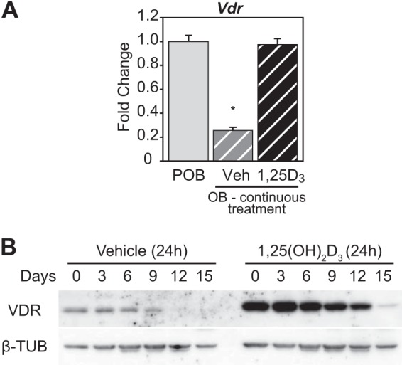
VDR expression is suppressed in OBs. A, POB cells differentiated for 15 days to OB cells were treated with 10−7 m 1,25(OH)2D3 (1,25D3) or Veh every 3 days continuously during culture. RT-qPCR was performed on RNA isolated from the POB and OB cells for Vdr levels. Values are relative quantitation (RQ) normalized to β-actin levels with fold change set to 1 for the POB sample (POB, gray; OB Veh, gray striped; OB 1,25(OH)2D3, black striped). Samples completed in biological triplicate ± S.E. *, p < 0.05 compared with POB sample. B, Western blot of VDR and β-tubulin (β-TUB) proteins from whole cell lysates collected every 3 days during differentiation from POB to OB (15 days). The cells were treated with vehicle or 10−7 m 1,25(OH)2D3 for 24 h prior to isolation.