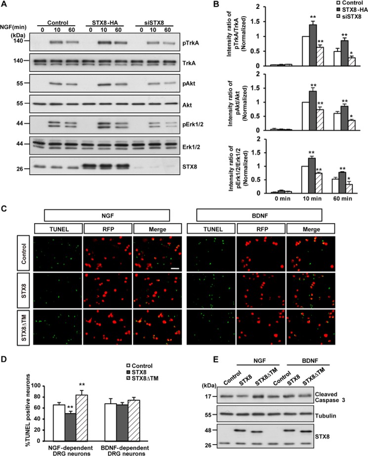
FIGURE 6.
STX8 regulates NGF-induced signal transduction and the survival of NGF-dependent DRG neurons. A, 615 cells transfected with STX8-HA, siSTX8, or control vector construct by electroporation were serum-starved overnight and treated with NGF (50 ng/ml) for 10 or 60 min. Immunoblotting was used to detect the phosphorylation of TrkA, Akt, and ERK1/2. B, quantification of TrkA, Akt, and ERK1/2 activation by the ratios of phospho-TrkA/Akt/ERK1/2 versus the total corresponding protein. The data are shown as the means ± S.E. (n = 3, *, p < 0.05; **, p < 0.01 versus their corresponding controls, one-way ANOVA). C, DRG neurons cultured for 24 h were infected with lentiviruses expressing RFP, STX8-RFP, or STX8ΔTM-RFP in the presence of NGF (50 ng/ml) or BDNF (50 ng/ml) for 7 days. After the neurons were switched to media containing 1.25 ng/ml NGF or BDNF for 24 h, TUNEL staining was performed according to the manufacturer's instructions. Scale bar, 100 μm. D, quantification of the percentage of apoptotic neurons. The data are shown as the mean ± S.E. (n = 3, **, p < 0.01 versus their corresponding controls, one-way ANOVA). E, DRG neurons treated as in C were lysed and immunoblotted to detect cleaved caspase-3.

