FIGURE 8.
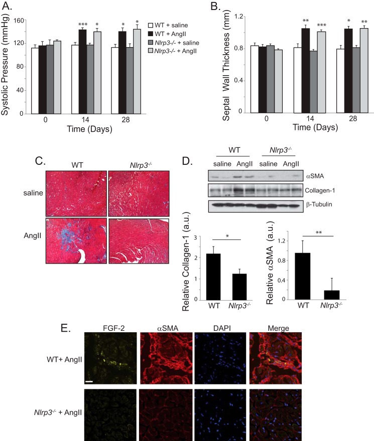
NLRP3 regulates AngII-induced cardiac fibrosis in vivo. A and B, systolic blood pressure and septal wall thickness from WT and Nlrp3−/− mice treated with saline (n = 4) or AngII (1.5 mg/kg/day, n = 7 WT, n = 8 Nlrp3−/−). *, p < 0.05; **, p < 0.01; ***, p < 0.001. C, Masson trichrome stain of left ventricular tissue taken at 28 days following saline or AngII infusion in WT and Nlrp3−/− mice. D, representative immunoblot and semiquantitative analysis for αSMA and collagen 1 in ventricular cardiac tissue taken from WT and Nlrp3−/− mice at 28 days following AngII or saline (n = 7 WT, n = 8 Nlrp3−/−). a.u., arbitrary units. *, p < 0.05; **, p < 0.01. E, confocal fluorescent immunohistochemistry showing localization of αSMA and FGF-2 in ventricular tissue from AngII-treated WT and Nlrp3−/− mice. Scale bar = 20 μm.

