FIGURE 6.

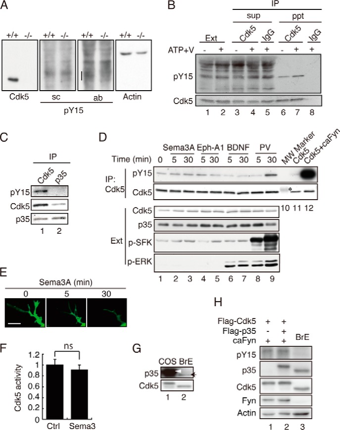
Tyr-15 phosphorylation of Cdk5 in neurons treated with Sema3A, Ephrin-A1, or BDNF. A, immunoblots of neuronal lysates with anti-phospho-Tyr-15 Cdk5 (pY15). Mouse cortical neurons prepared from wild-type (+/+) or Cdk5 knockout (−/−) mouse brains were immunoblotted with antibodies to Cdk5 C8, anti-phospho-Tyr-15 of Cdk5 (sc) and (ab), and actin. B, an immunoblot of anti-Cdk5 C8 immunoprecipitates with anti-phospho-Tyr-15 (sc). Cdk5 was immunoprecipitated (IP) from the extract (Ext) of mouse brains at embryonic day18.5, which was treated with Na3VO4 (V) and ATP. The supernatant (sup) or immunoprecipitate (ppt) was immunoblotted with anti-Cdk5 C8 or control IgG, followed by immunoblotting with anti-phospho-Tyr-15 Cdk5 (sc) (top panel) or anti-Cdk5 C8 (bottom panel). C, phospho-Cdk5 at Tyr-15 did not coimmunoprecipitate with anti-p35. Cultured neuronal lysates were immunoprecipitated with anti-Cdk5 C8 or anti-p35 C19, and Cdk5 phosphorylated at Tyr-15 was detected with anti-phospho-Tyr-15 Cdk5 (sc). D, Tyr-15 phosphorylation in neurons treated with Sema3A, Ephrin-A1, or BDNF. Mouse cortical neurons were treated with Sema3A, Ephrin-A1 (Eph-A1), BDNF, or 100 μm pervanadate (PV) for the indicated times. Cell lysates were immunoblotted with antibodies to Cdk5 (third panel), p35 (fourth panel), anti-phospho-SFK (fifth panel), or phospho-Thr-202/Tyr-204 of ERK (p-MAPK, sixth panel). Phosphorylation of Tyr-15 on Cdk5 was detected with anti-phospho-Tyr-15 Cdk5 (sc) (first panel) after immunoprecipitation with anti-Cdk5 C8 (second panel). Lane 10 is a molecular weight (MW) marker. The asterisk indicates carbonic anhydrase at 32.2 kDa. Lanes 11 and 12 are Cdk5 coexpressed with or without caFyn in HEK293 cells for reference. E, the effect of Sema3A on neurite retraction. Neurites of mouse cortical neurons expressing EGFP were observed by time-lapse imaging at intervals of 5 min after addition of Sema3A. Scale bar = 5 μm. F, kinase activity of Cdk5 after Sema3A treatment. Cdk5 was prepared from cultured neurons treated with Sema3A for 30 min by immunoprecipitation, and its kinase activity was measured by phosphorylation of histone H1. Data are mean ± S.E. (n = 3). ns, not significant; Student's t test. Ctrl, control. G, the protein ratio of p35 and Cdk5 in COS-7 cells and mouse brain extract. FLAG-Cdk5 and FLAG-p35 were transfected into COS-7 cells using 1 μg of plasmid for each of Cdk5 and p35, the same as in Fig. 2. Immunoblotting was performed by adjusting Cdk5 approximately (bottom panel). FLAG-p35 expressed in COS-7 cells is indicated by an arrowhead, and p35 in the brain extract (BrE) is indicated by an arrow (top panel). H, the effect of p35 at the low expression levels (similar to the brain extract) on Tyr-15 phosphorylation of Cdk5. COS-7 cells were transfected with FLAG-Cdk5 and caFyn with or without FLAG-p35 as shown in Fig. 2A, except for the reduced plasmid amount of p35 (1/40). Phosphorylation of Tyr-15 in Cdk5 was detected by immunoblotting with antibodies to phospho-Tyr-15 of Cdk5 (ab). Blotting of Cdk5, p35, Fyn, and actin is also shown.
