FIGURE 5.

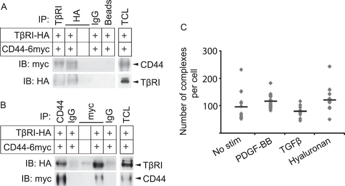
TβRI and CD44 form a ligand noninducible complex. A and B, Cos1 cells were transiently transfected with HA-tagged TβRI, 6-Myc-tagged CD44, or corresponding empty tagged vectors. After 48 h, lysates were immunoprecipitated (IP) with antibodies against TβRI or HA (A), CD44 or Myc (B), corresponding IgG isotype controls, or beads alone; proteins were separated by SDS-PAGE. Total cell lysates (TCL) were run in parallel. Immunoblotting (IB) was performed with antibodies against CD44 and TβRI. C, BJ-hTERT foreskin fibroblasts were grown in 8-well chambers, starved, and stimulated (stim) for 7 min with PDGF-BB (20 ng/ml), 2 h with hyaluronan (200 μg/ml), or 1 h with TGFβ (1 ng/ml). Cells were fixed, and PLA was performed with mouse anti-CD44 and rabbit anti-TβRI antibodies, followed by anti-mouse and anti-rabbit PLA probes conjugated with priming and nonpriming oligonucleotides. F-actin was stained with FITC-conjugated phalloidin. Individual protein-protein interactions were visualized by fluorescence microscope as red dots. PLA signals per cell were quantified using Duolink Image Tool according to the manufacturer's instructions. Average values are indicated in the graph by horizontal lines. A representative experiment out of three is shown.
