
Fig. 2.
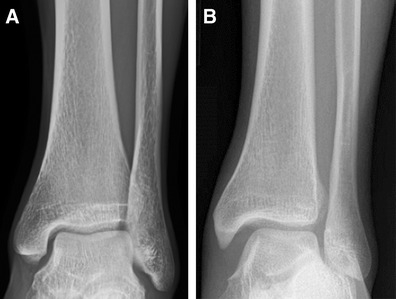
Anteroposterior radiographs of a normal ankle (a) and an ankle with syndesmotic injury (b). Note the widened medial clear space, loss of tibiofibular overlap, and widening of the distal tibiofibular syndesmosis
Official websites use .gov
A
.gov website belongs to an official
government organization in the United States.
Secure .gov websites use HTTPS
A lock (
) or https:// means you've safely
connected to the .gov website. Share sensitive
information only on official, secure websites.
Anteroposterior radiographs of a normal ankle (a) and an ankle with syndesmotic injury (b). Note the widened medial clear space, loss of tibiofibular overlap, and widening of the distal tibiofibular syndesmosis