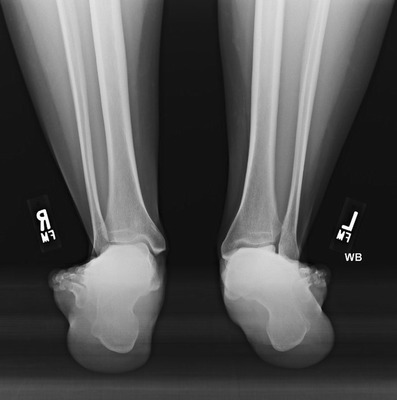
Fig. 5.

A weight-bearing, hindfoot alignment view of both feet demonstrates bilateral hindfoot valgus deformity as evidenced by the lateral and valgus position of the calcaneus with respect to the axis of the tibia. The left (labeled “L”) is much more severe than the right (labeled “R”)
