
FIG. 7.
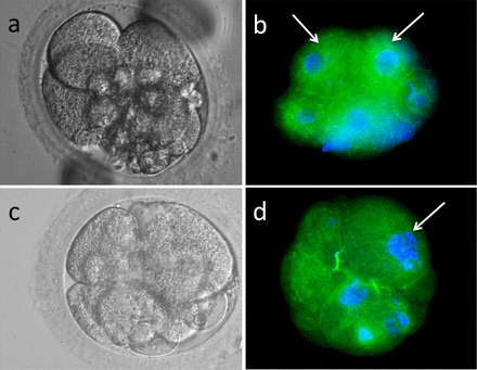
Fluorescence staining of four-cell rhesus embryos from XXO-treated sperm treatment group stained for α-tubulin (monoclonal anti-α-tubulin-FITC antibody) and Hoescht 33342 nuclear stain. a) Hoffman modulation contrast image of a four-cell embryo. b) Fluorescence image of same embryo shown in a. Note severe cellular fragmentation of DNA-containing fragmented cells; two blastomeres appear normal (arrows). c) Hoffman modulation contrast image of a four-cell fragmenting embryo demonstrating cellular debris and uneven-sized blastomeres. d) Fluorescence image of same embryo demonstrating several blastomeres without detectable DNA, fragmenting nuclei (arrow), and cell fragments containing DNA. Original magnification ×400.