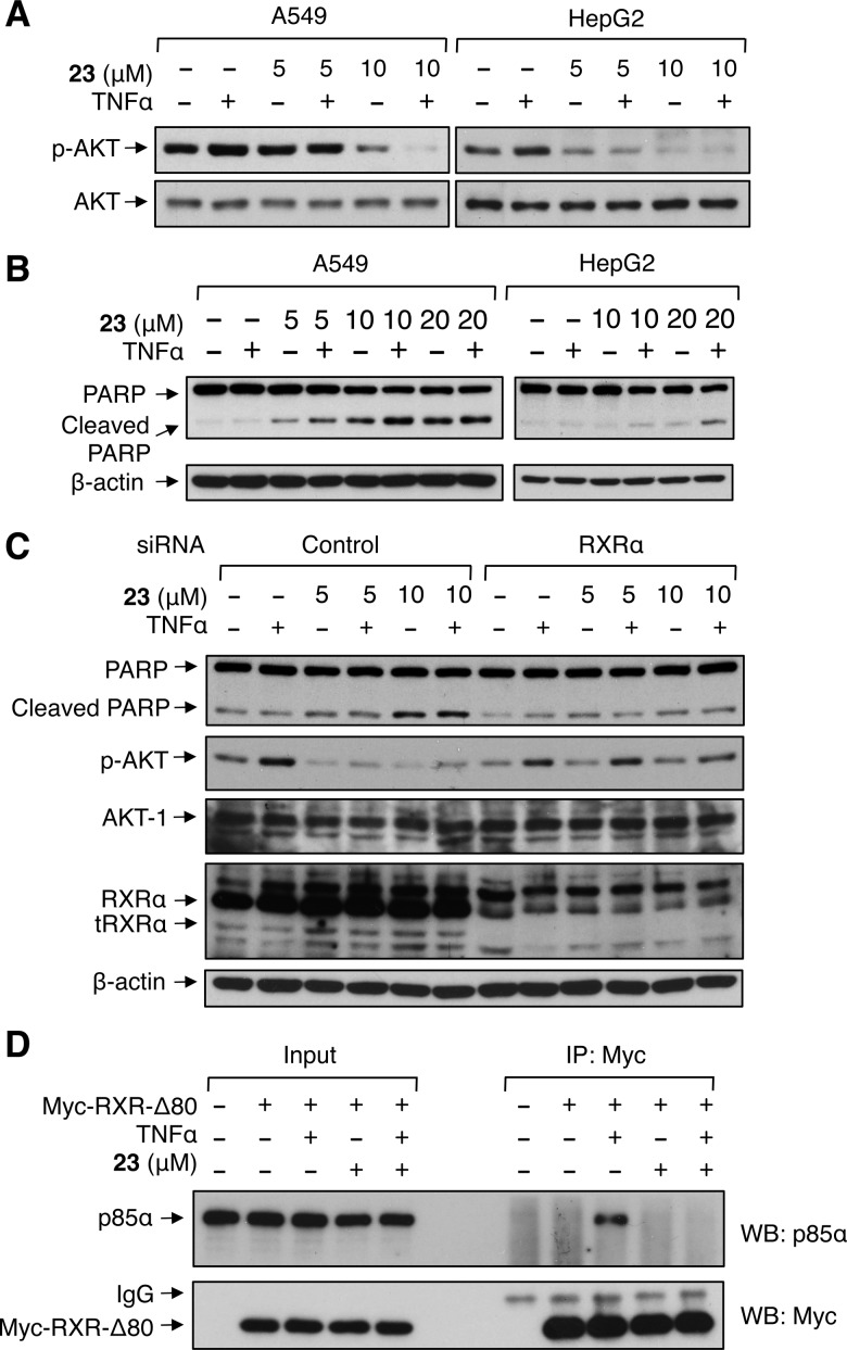
Figure 4.
Biological evaluation of 23. (A,B) Inhibition of AKT activation (A) and induction of apoptosis (B). Cells were pretreated with 23 for 24 h before being exposed to TNFα (20 ng/mL) for an additional 30 min. Lysates prepared were analyzed by Western blotting for AKT activation (A) or PARP cleavage (B). (C) RXRα-dependent effects of 23. A549 cells transfected with RXRα siRNA or control siRNA for 48 h were treated with 23 for 24 h before being exposed to TNFα (20 ng/mL) for an additional 30 min. Lysates prepared were analyzed by Western blotting. (D) Inhibition of p85α interaction with tRXRα by 23. A549 cells transfected with myc-RXRα-Δ80 expression vector were analyzed for their interaction with endogenous p85α by coimmunoprecipitation assay using anti-Myc antibody. Immunoprecipitates were analyzed by Western blotting for the presence of p85α and Myc-RXRα-Δ80. One of three to five similar experiments is shown.

