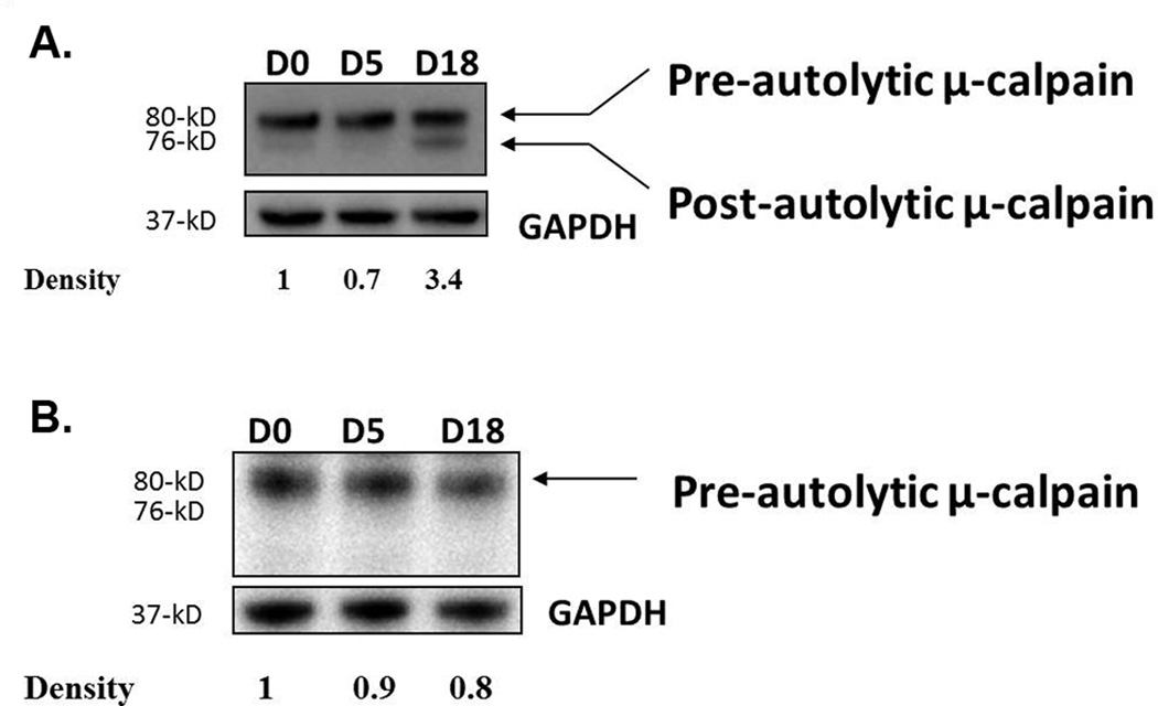
Figure 2.
Activation of µ-calpain following infection. WT (a) and SCID (b) mice were infected with the GS strain of G. duodenalis and jejunal homogenates were tested for activated µ-calpain by Western blot using an antibody capable of detecting both active and inactive forms of µ-calpain. GAPDH was used as a loading control. Each figure is representative of 4 mice/time point.

