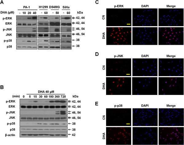
Figure 2.

DHA activates MAPKs. (A) DHA induces MAPKs activation. PA-1, H1299, D54MG and SiHa cell lines were treated with the indicated doses of DHA for and 24 h (12 h in case of PA-1 cells). Then, protein lysates were separated and immunoblotted with antibodies against conventional MAPKs. (B) Expression patterns of conventional MAPKs in response to DHA over time. PA-1 cells treated with 40 μM DHA for the indicated time periods were subjected to immunoblotting for MAPKs. (C-E) Nuclear accumulation of phospho-ERK, -JNK, and -p38 in PA-1 cells after DHA exposure. PA-1 cancer cells were incubated for 6 h with or without 40 μM DHA. Then, cells were stained with antibodies against phospho-ERK (C), phospho-JNK (D) and phospho-p38 (E) and analyzed by immunoflurescence. Scale bars, 50 μm.
