
Figure 4. Brown adipose tissue and respiratory muscle activity during the thermoneutral and cold exposure experiment. A, D.
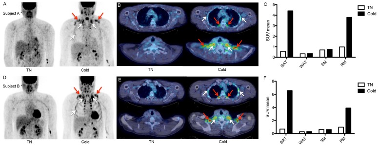
) PET images during thermoneutral (left) and cold (right) conditions showing [18F]FDG-uptake e in brown adipose tissue (BAT; red arrows) and respiratory muscles (RM; white arrows). B, E) Transaxial slices of subject A (5 mm thick) of thoracic area (upper) and supraclavicular area (lower) demonstrating BAT activity (red arrows) and RM activity (white arrows). C, F) [18F]FDG-uptake (SUVmean) in BAT, white adipose tissue (WAT), skeletal muscle (SM), and respiratory muscles (RM) during thermoneutral and cold conditions.