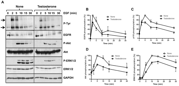
Figure 3. Effect of testosterone treatment on ligand-dependent activation of the EGFR and downstream signaling pathways in smooth muscle vascular cells.
Serum starved SMVC were incubated with 10% (w/v) trichloroacetic acid. The samples were processed by Western blots using the indicated antibodies to determine the phosphorylation levels of the EGFR, p115, Akt, and ERK1/2, and the total levels of EGFR Akt, ERK1/2 and GAPDH as described in Materials and Methods. The total EGFR level decreased after EGF stimulation due to the expected proteolytic processing of the receptor after ligand-dependent internalization. The total Akt, ERK1/2 and GAPDH levels were used as loading controls. The intensity of the bands was measured densitometrically and the signal of the different phosphoproteins was corrected using appropriate loading controls. The photograph (A) shows typical Western blots of the proteins. The top and bottom arrows point to the phosphorylated EGFR and p115, respectively. The plots (B–E) present the mean ± SEM phosphorylation of the EGFR (n = 4) (B), p115 (n = 5) (C), Akt (n = 6) (D), and ERK1/2 (n = 6) (E) from a set of experiments similar to those shown in A.

