
Figure 5. ZFP36L1 mediates degradation of a 3′ UTR BCL2 ARE.
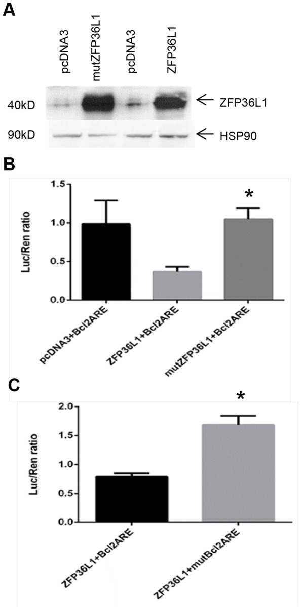
(A) Transfection of ZFP36L1 or mutant ZFP36L1 into HEK293T cells results in high level ZFP36L1 expression as shown by Western blot analysis. Very low/absent levels of ZFP36L1 are found in empty vector (pcDNA3) transfected HEK293T cells. (B) ZFP36L1 interacts with the BCL2 ARE and mediates BCL2 ARE degradation. BCL2 3′ UTR luciferase reporter assay showing reduced luciferase values in HEK293T cells transfected with ZFP36L1 and BCL2 ARE compared to values obtained by co-transfection of BCL2 ARE with either an empty vector (pcDNA3) or a zinc finger mutant ZFP36L1. The results show mean ±SEM for three independent experiments. p<0.05 as determined by student T test. (C) BCL2 3′ UTR Luciferase reporter assay showing that ZFP36L1 requires the BCL2 ARE core AU-rich element to effectively reduce luciferase levels, whereas a BCl2 ARE construct with the core AU-rich element removed is much less affected. The results show mean ±SEM for three independent experiments, p<0.05 as determined by student T test.