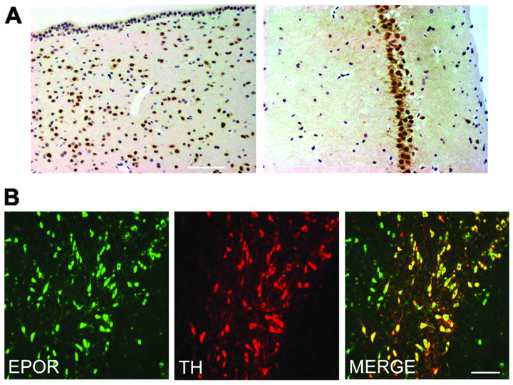
Figure 3.
Immunohistochemical analysis of erythropoietin receptor (EPOR) expression in brain sections from rat. (A) Staining of the periventricular zone and hippocampus of rat showed neuron staining for EPOR. (B) Double-staining for tyrosine hydroxylase (TH) showed that TH-immunoreactivity (TH-IR) dopaminergic neurons in the SNpc also express EPOR. Scale bar, 100 μm.

