
Figure 6.
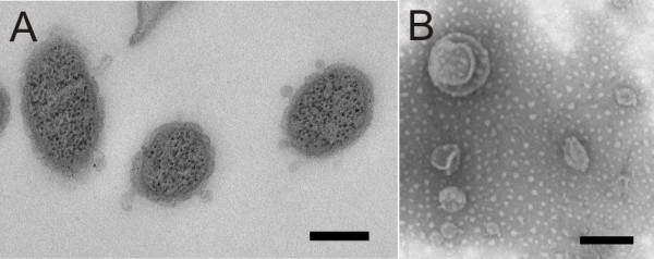
OMVs of Rhizoid morphotype of F. columnare B067. Panel A: Outer membrane vesicles visualised under TEM from the thin sections of the cells of the F. columnare; and Panel B: Purified vesicles under TEM. In the thin sections, the size of the observed vesicles was under 100 nm, whereas the purified vesicles ranged from 60 to 350 nm. The scale bar was 200 nm.