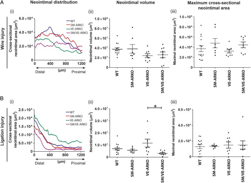
Figure 7.
Effect of vascular-specific AR ablation on neointimal lesion formation. Lesion formation following (A) wire-induced injury (n = 7–14) or (B) ligation-induced injury (n = 6–14) was determined by optical projection tomography (OPT) and histology. Panels A(i) and B(i) show mean neointimal lesion volumes for each genotype; error bars have been omitted for clarity. Panels A(ii and iii) an B(ii and iii) show individual data points from each animal with lines and error bars indicating mean ± SEM. Vascular AR deletion had no effect on lesion formation in response to wire-induced injury. Selective deletion of AR from ECs produced a small increase in neointimal lesion volume following ligation injury (*P < 0.05 by one-way ANOVA plus Bonferroni post-hoc test) but did not alter maximal cross-sectional narrowing. (WT = wild-type litter mates carrying floxed-AR; SM-ARKO = AR ablated in SMC, VE-ARKO = AR ablated in EC, SM/VE-ARKO = AR ablated in both EC and SMC.)

