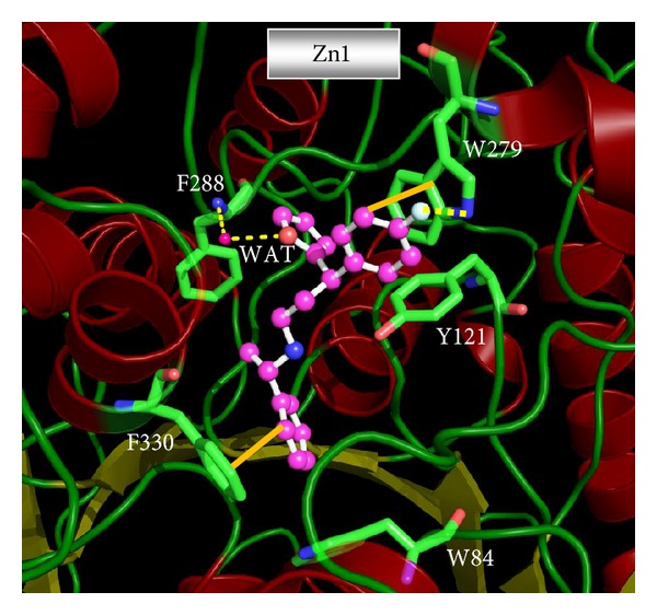
Figure 9.

Molecular docking derived binding pose of the VS lead compounds Zn1 in the dual binding site (CS and PAS) of AChE enzyme. The inhibitor is shown as ball and stick model in the surface representation of the enzyme. Water compounds are shown as red dotted spheres. GOLD software was used to derive the binding mode and the picture was generated from PyMOL software.
