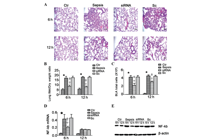
Figure 3.
Histological examination of the lung sections at 6 and 12 h after different treatments of mice with CLP-induced ALI. (A) Lung tissue from the control, sepsis, siRNA and Sc groups at 6 and 12 h after surgery (hematoxylin & eosin staining; magnification, ×200); (B) wet/dry ratio of lungs from the control, sepsis, siRNA and Sc groups at 6 and 12 h after surgery; (C) total cell number in the BAL fluid of mice from different groups; (D and E) levels of NF-κB mRNA and protein in the lung tissue of mice from different groups. Data are expressed as the mean + standard error (n=10 per group). *P<0.05 compared with the Ctr group; †P<0.05 compared with the sepsis group; ‡P<0.05 compared with the Sc group. siRNA, small interfering RNA; CLP, cecal ligation and puncture; ALI, acute lung injury; Sc, scrambled control group; Ctr, control group; NF-κB, nuclear factor κB; BAL, bronchoalveolar lavage.

