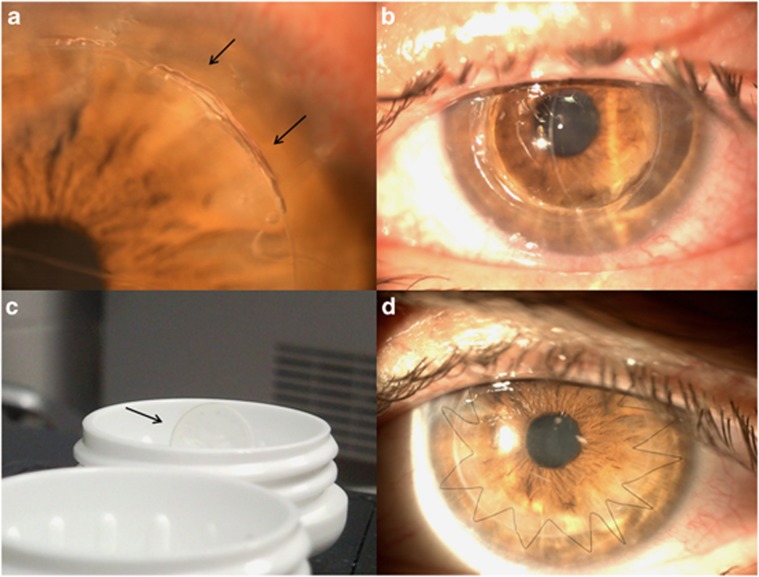
Figure 1.
(a) Slit lamp photograph shows a surgical wound dehiscence located at 1–2 o'clock hours after suture removal. (b) The clinical appearance seen through slit lamp photograph reveals the absence of the whole donor graft and the DM exposed. Note the anterior chamber is deep and formed. (c) The contact lens case shows the graft inside. Slit lamp photograph 1 month after the second DALK is shown in (d).

