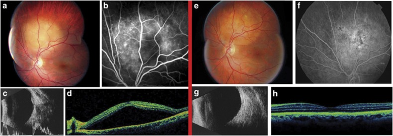
Figure 1.
Regression of choroidal metastasis after intravitreal bevacizumab (Case A). Pre-treatment (left images): (a) Fundus photograph shows yellow mass in the superior sector close to the optic disc, (b) FA demonstrates hypofluorescence due to masking effect with signs of leakage, (c) B-scan echography shows a medium-high reflective choroidal mass above the optic disc, (d) OCT demonstrates serous detachment of the neuroepithelium. Post-treatment (right images): (e) Fundus photograph shows regression of the choroidal mass, (f) FA demonstrates pigmentary changes with hypo-hyperfluorescent areas and signs of scarring due to flattening of the mass, (g) B-scan echography shows regression of the mass, (h) OCT demonstrates resolution of the serous detachment.

