
Figure 11.
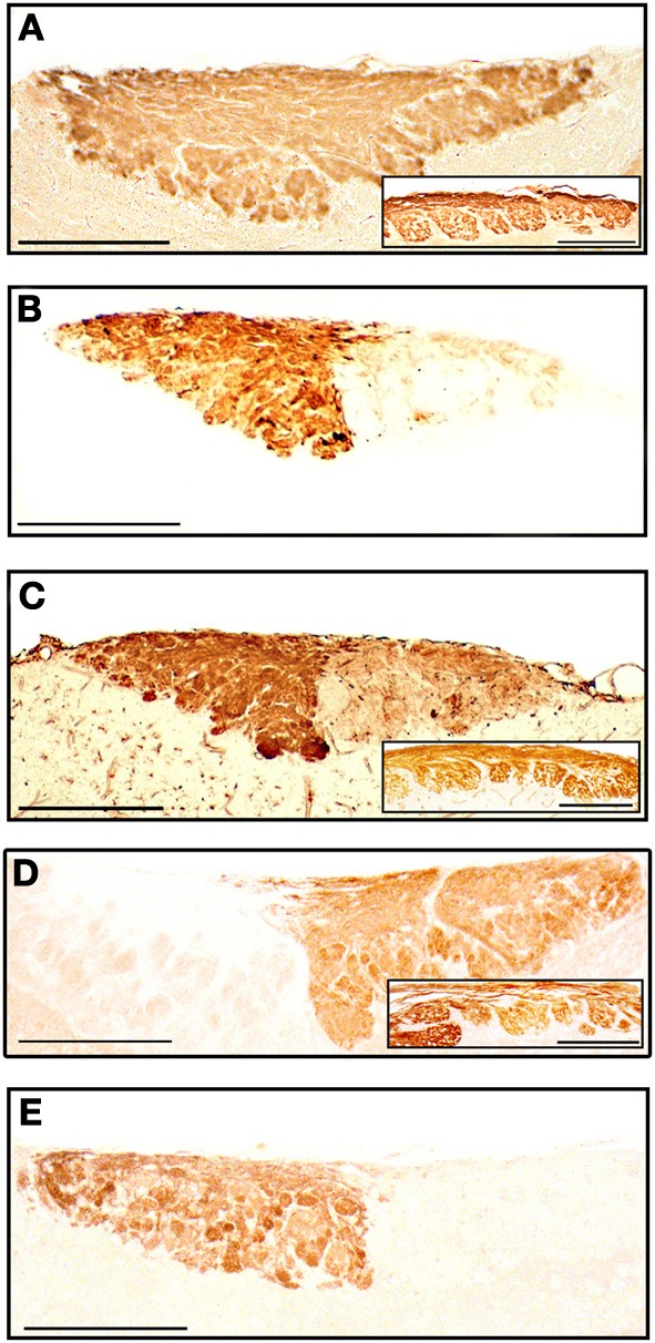
Parasagittal sections of the olfactory bulb through the AOB (left anterior, right posterior) stained with anti-OMP (A), UEA-I (B), LEA (C), anti-Gα0 (D) and anti-Gαi2 (E). Insets show the nervous and glomerular layers of the MOB. Scale bars: (A) 250 μm; (B–E) and insets, 200 μm.