
Figure 4.
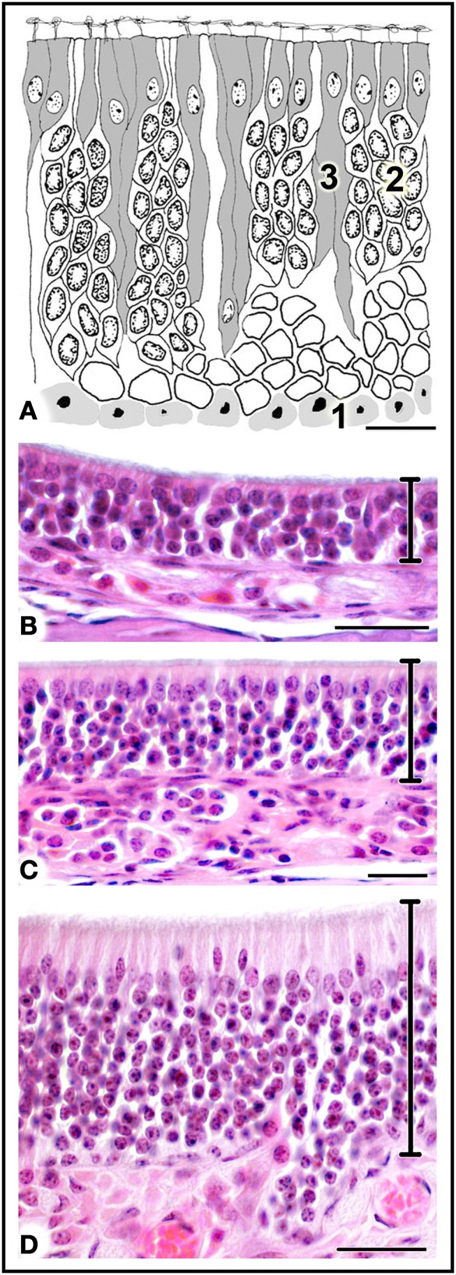
(A) Diagrammatic reconstruction of main olfactory epithelium, showing basal cells (1), mature neurons (2) and supporting cells (3) (modified after Graziadei, 1971). (B,C) Haematoxylin-eosin-stained sections of areas of epithelium with different thicknesses (see text). Scale bars: (A) 10 μm; (B) 20 μm; (C,D) 25 μm.