Figure 6.

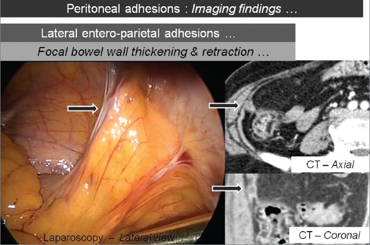
Axial and coronal CT images showing the focal thickening and retraction of bowel wall with corresponding laparoscopic image (arrows), which suggests lateral entero-parietal adhesions

Axial and coronal CT images showing the focal thickening and retraction of bowel wall with corresponding laparoscopic image (arrows), which suggests lateral entero-parietal adhesions