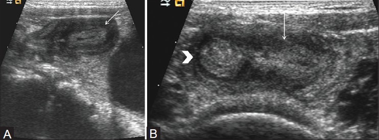
Figure 10(A, B).

Meckel's diverticulum. (A) Longitudinal view of right lower quadrant shows oval structure (arrow) with central echogenic mucosa and peripheral hypoechoic muscle. (B) Longitudinal view of right lower quadrant shows connection (arrow) of this oval hypoechoic structure with distal ileum (arrow head) Normal appendix was seen separate from the lesion. On surgery, inflamed Meckel's diverticulum was identified
