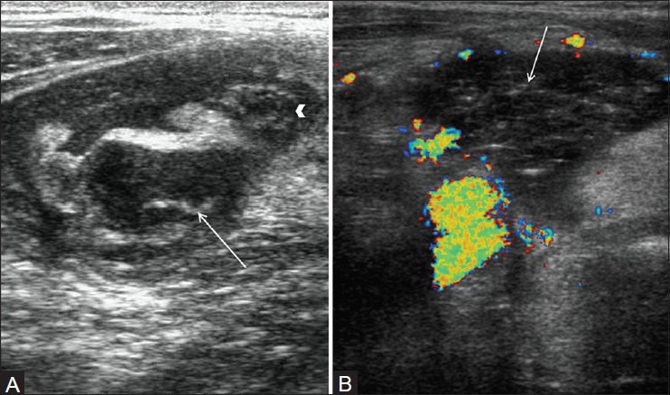
Figure 5(A, B).

Perforated appendicitis. (A) Short axis view through right lower quadrant demonstrates enlarged appendix (arrowhead) with surrounding collection in right iliac fossa and appendicolith (arrow) in the fluid collection consistent with perforated appendix. (B) Color Doppler image in another patient shows complex collection (arrow) in right iliac fossa without internal vascularity consistent with abscess secondary to appendicular perforation
