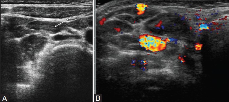
Figure 7(A, B).

Mesenteric lymphadenopathy. (A) Longitudinal view through the right lower quadrant using graded-compression sonography shows several enlarged, oval-shaped, mesenteric lymph node with echogenic central hilum (arrow). (B) Color Doppler scan in another patient shows an enlarged node (arrow) with central flow
