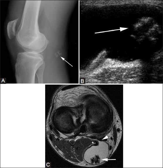
Figure 1(A-C).

Popliteal cyst. A 60-year-old female with knee pain. (A) Lateral knee radiograph demonstrates coarse calcifications (arrow) in the popliteal fossa. (B) Gray scale USG image at the level of popliteal fossa demonstrates a cystic lesion containing echogenic calcifications (arrow) with posterior acoustic shadowing. (C) Axial T2W image through right knee demonstrates the hyperintense popliteal cyst fluid arising between semimembranosus (arrowhead) tendon and medial head of gastrocnemius (asterisk), with hypointense loose bodies (arrow) layering dependently
