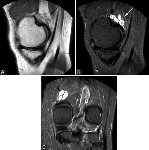
Figure 10(A-C).

Gastrocnemius ganglion. A 45-year-old male with knee pain. Sagittal PDW image (A) demonstrates lobulated lesion (arrows) at insertion of medial head of gastrocnemius that is hyperintense on sagittal (B) and coronal (C) T2W fat-saturated images
