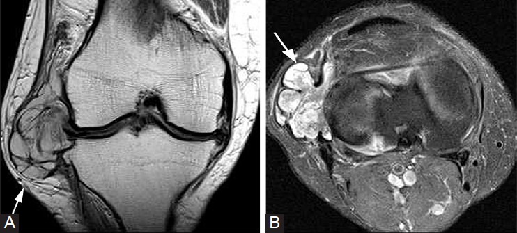
Figure 14(A, B).

Meniscal cyst. A 42-year-old female with knee pain and palpable lateral mass. (A) Coronal PDW image of the right knee demonstrates a multilobulated cyst (arrow) with internal septations, which arises from tear of body of lateral meniscus. (B) T2W fat-saturated image demonstrates cystic nature of this lesion (arrow), which did not enhance following contrast administration (not shown)
