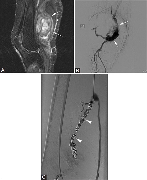
Figure 18(A-C).

Popliteal artery aneurysm. A 62-year-old male with pulsatile mass in popliteal fossa. (A) Sagittal T2W fat-saturated image demonstrates heterogeneous mass in the region of popliteal fossa (arrows). (B) Digital subtraction angiography shows partial thrombosis of large popliteal fossa aneurysm (arrows), which was subsequently embolized with coils (arrowheads) (C)
