Figure 19(A, B).

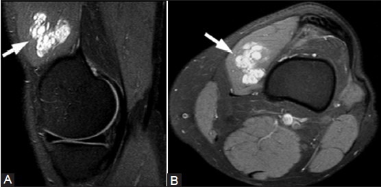
Intramuscular hemangioma. A 54-year-old female with knee mass and pain. (A) Sagittal T2W fat-saturated and (B) axial T1W fat-saturated post-contrast images show a lobulated enhancing cystic mass (arrows) in the vastus medialis

Intramuscular hemangioma. A 54-year-old female with knee mass and pain. (A) Sagittal T2W fat-saturated and (B) axial T1W fat-saturated post-contrast images show a lobulated enhancing cystic mass (arrows) in the vastus medialis