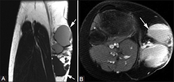
Figure 20(A, B).

Lymphatic malformation. A 23-year-old female with palpable medial knee mass. (A) Coronal T1W image shows multilobulated cystic lesion (arrows) in the medial aspect of the knee that is isointense and hyperintense to muscle. (B) Axial fat-suppressed T2W image shows fluid-fluid levels (arrow) in this multicystic lesion
