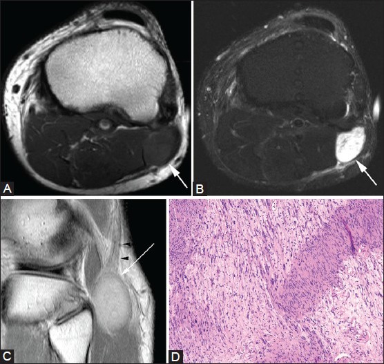
Figure 23(A-D).

Schwannoma. A 38-year-old male with knee pain. Axial (A) T1W and (B) T2W fat-suppressed images show T2-hyperintense lesion (arrows) in the lateral soft tissue of the knee. (C) Coronal PDW image confirms that the mass (arrow) arises from common peroneal nerve (arrowheads). (D) Photomicrograph [hematoxylin and eosin (H and E), ×400] shows alternating hypocellular and hypercellular spindle cells with Verocay bodies and cytologically bland spindle cells with plump nuclei, consistent with a schwannoma
