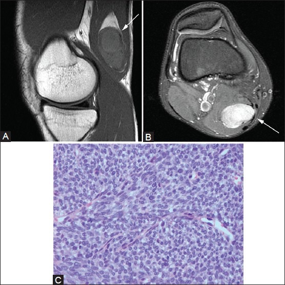
Figure 24(A-C).

Synovial sarcoma. A 23-year-old male with palpable mass. (A) Sagittal T1W image demonstrates hypointense oblong mass in semimembranosus muscle (arrow), which has solid internal enhancement (arrow) on post-contrast T1W fat-saturated image (B). (C) Photomicrograph (H and E, ×200) shows a highly cellular spindle cell neoplasm composed of densely packed cells with scant cytoplasm. The tumor is characterized by its lack of nuclear pleomorphism and a mitotic rate that is lower than expected for such a cellular tumor, consistent with a synovial sarcoma
