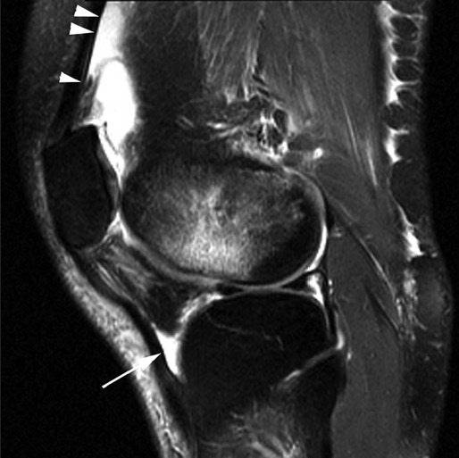
Figure 6.

Infrapatellar bursitis. A 28-year-old male with anterior knee pain. Sagittal fat-saturated T2W image shows triangular pocket of fluid (arrow) between distal patellar tendon and anterior tibia. There is also bone contusion in lateral femoral condyle and fluid in suprapatellar recess (arrowheads), resulting from transient lateral patellar dislocation
