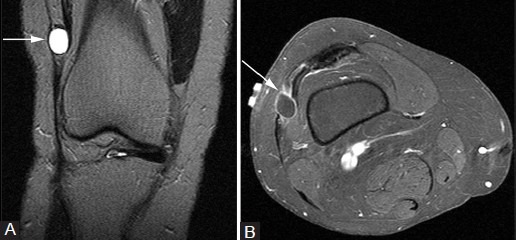
Figure 7(A and B).

Iliotibial band cyst. A 46-year-old man with palpable abnormality. (A) Coronal T2W fat-saturated image demonstrates well-circumscribed hyperintense cyst (arrows) abutting the iliotibial band, which does not enhance internally following contrast administration on the post-contrast T1W fat-saturated image (B)
