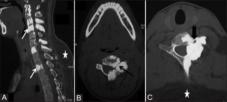
Figure 2(A-C).

(A) Sagittal reformatted CT scan image showing hyperostosis involving cervical and dorsal vertebrae. (B and C) Axial CT scan images showing narrowing of spinal canal, neural foraminae (black arrow), and left foramen transversarium (arrowhead). Incidental note is made of lipoma (asterisk) at back
